Free standard shipping for orders over $340.

Blog

Uncategorized

Glucosamine For Therapeutic Use, Mainly as an Antiarthritic Agent

Glucosamine (chemically 2-amino-2-deoxyglucose), commonly known as chitosamine is currrently recognized as a safe and effective antiarthritic agent1. It is found in mucopolysaccharides and mucoproteins, which form the frame work of vertebrate connective tissues. It occurs extensively as chitin in the exoskeleton of invertebrates, including arthropods and marine organisms. Microbes such as yeast and other fungi also contain chitin. Chitin is a biopolymer made up of units of N-acetyl-glucosamine. Glucosamine can be isolated from chitin or prepared synthetically. For pharmaceutical use as an antiarthritic agent, glucosamine is supplied in the form of products such as glucosamine sulfate.

Glucosamine sulfate is the preferred form of glucosamine for therapeutic use, mainly as an antiarthritic agent. It is an effective means of providing glucosamine orally, as a building block for the regeneration of cartilage glycosaminoglycans, lost during the progression of osteoarthritis.

Chemistry of glucosamine sulfate:

Each unit of glucosamine sulfate contains two molecules of glucosamine as below:

In vivo, glucosamine sulfate breaks down into glucosamine and sulfate ions.

Pharmacological action:

Cartilage glycosaminoglycans are formed from glucosamine. Glucosamine is biochemically generated in the body from fructose-6-phosphate, a product of glycolysis. The glucosamine formed is converted to galactosamine, which is then incorporated into glycosaminoglycans and glycoproteins, the building blocks of cartilage tissue.

During the progression of osteoarthritis, there is substantial degeneration of cartilage glycosaminoglycans. The availability of glucosamine, a critical intermediate in the synthesis of mucopolysaccharides in cartilage tissue, is rate limiting for proteoglycan production. Glucosamine administration could therefore be expected to induce the formation of glycosaminoglycans, thereby restoring the depleted glycosaminoglycans and proteoglycans. The anti-inflammatory activity of glucosamine sulfate have also been reported by several researchers. The following effects of glucosamine sulfate have been proven in several detailed studies:

  1. Biosynthesis of glycosaminoglycans
  2. Stimulation of the synthesis of proteoglycans
  3. Antireactive action


Preclinical studies:

Preclinical studies were directed at understanding the mechanism of action of glucosamine sulfate as an antiarthritic agent and its safety during subacute or chronic administration.

  1. The antireactive properties were studied in rats and mice. Inflammation was induced in these animal models with several non specific agents (carrageenin, dextran, formalin and acetic acid) and specific agents (serotonin, bradykinin and histamine). The spectrum of activity was shown to be restricted to the inflammatory processes provoked by non specific agents.
  2. Glucosamine was shown to have no analgesic activity but was able to reduce the generation of superoxide radicals by macrophages and inhibit lysosomal enzymes.

The antiarthritic effects of glucosamine sulfate are best described as being linked to its antireactive properties. The mechanism of action is therefore thought to be different from that of conventional non steroidal antiinflamatory drugs. Unlike conventional NSAIDs, Glucosamine sulfate acts by stimulating the synthesis of proteoglycans which stabilize the cell membranes and the intercellular substance. In view of this mechanism of action, glucosamine sulfate can be said to have antireactive rather than antiinflammatory properties. Glucosamine sulfate has cyclooxygenase-independent antireactive properties and no analgesic action.

The comparative protective effects of glucosamine sulfate and indomethacin are shown in Table 1:

Table 1 : Inhibitory concentrations on mediators of inflammation (concentrations able to inhibit the investigated mediator by 25% (IC25) or by 50% (IC50) 1


Model

Glucosamine sulfate

IC (mmol/l)

Indomethacin

IC (mmol/l)

Rat : proteolytic enzymes in inflamed paw IC25 > 3 IC25 0.33
Rat : lysosomal enzymes of

the liver

IC25 3 IC25 0.27
Superoxide generation IC25 > 3 IC25 0.07
Inhibition of prostaglandin synthesis

¾ Stimulated by arachidonic acid

IC50 > 660 IC50 0.50
¾ Stimulated by histamine IC50 > 660 IC50 0.53

From the above table, it may seem that glucosamine sulfate shows a very low potency as compared to indometacin. However, glucosamine sulfate has very low toxicity making it more suitable for long term administration. The LD50 values for oral administration of glucosamine sulfate, indometacin and acetyl salicylic acid are shown in Table 2:

Table 2 : LD50 values by oral route

Substance LD50 values (mg/kg) by oral route
Rat Mouse
Glucosamine > 8000 > 8000
Indometacin 12.3 12.8
Acetyl salicylic acid 1435 1050


The antireactive activity of glucosamine sulfate was tested in rats8 with experimental models such as:

a) sponge granuloma and croton oil induced granuloma (subacute inflammation)
b) kaolin arthritis (subacute mechanical arthritis)
c) Immunological reactive arthritis
d) Generalized inflammation (adjuvant arthritis)

Glucosamine sulfate was found to be effective in oral daily doses of 50 to 800 mg/kg. The potency of glucosamine sulfate in comparison with that of indometacin in the same tests was found to be 50-300 times lower. However, toxicity of indometacin in chronic toxicity experiments was found to be 1000 to 4000 times larger. The therapeutic margin with regard to prolonged treaments required for inflammatory disorders results 10-30 times more favorable for glucosamine sulfate than for indometacin. Based on these observations in animal models, glucosamine sulfate is evidently the drug of choice for prolonged treatment of inflammatory disorders8.

Additionally, the anti-viral properties and anti-cancer properties of glucosamine have been investigated and its inhibitory effects on a carcinoma have been demonstrated5. Currently, however, the focus is on the use of glucosamine sulfate in the management of rheumatic disorders.

Clinical studies:

  1. In one of the early double blind placebo controlled clinical studies , 80 patients found significant pain relief and improved motility with a daily dose of 1.5 g of glucosamine for 30 days. Histological examinations in these patients revealed restoration of healthy cartilage after treatment, lending credence to the fact that glucosamine sulfate helps build cartilage tissue7.
  2. A number of independent double blind studies performed over the last fifteen years indicate that glucosamine sulfate may be superior to some non-steroidal antiinflammatory drugs and placebos in the treatment of osteoarthritis5.
  3. Intramuscular glucosamine sulfate was found to be effective with negligible side effects in the treatment of osteoarthritis of the knee, in a randomized placebo-controlled double blind study2. The results of this study are summarized in Figure 1. The Lequesne index is a measure of the severity of the symptoms of osteoarthritis. A significant (p < 0.05) decrease in the index was observed for glucosamine sulfate as compared to placebo.
EFFICACY OF GLUCOSAMINE SULFATE (INTRAMUSCULAR, 400 mg, twice a week for six weeks) IN THE TREATMENT OF OSTEOARTHRITIS OF THE KNEE AS COMPARED TO PLACEBO
  1. Other similar studies revealed improved joint functions and reduced pain in patients treated with glucosamine as compared with those treated with a placebo7.
  2. One study compared the effects of glucosamine and ibuprofen in patients suffering from osteoarthritis of the knee. A 1.5 g oral daily dose of glucosamine for eight weeks was more effective than ibuprofen in reducing pain6.

Rationale behind the preferential use of Glucosamine sulfate over Glucosamine hydrochloride:

  1. Glucosamine sulfate occurs naturally in the human body and is almost devoid of toxicity, making it suitable for long term clinical use1.
  2. The chondrometabolic, antireactive and antiarthritic properties of this form have been investigated extensively and are supported by controlled clinical trials2.
  3. Pharmacokinetic studies revealed that close to 90% of orally administered glucosamine sulfate is absorbed3.
  4. Glucosamine sulfate, in vivo, breaks down into glucosamine and sulfate ions. Glucosamine hydrochloride, on the other hand, breaks down into glucosamine and free hydrochloric acid. The acid would increase gastric acidity and produce the associated disturbances in vivo.
  5. The sulfate ions produced from glucosamine sulfate, on the other hand, participate in chondrometabolic processes. They influence the incorporation of glucosamine and sulfate into the tissues. In a study with gastric mucosal segments, a sulfate content of 300 mmole was found to induce maximum incorporation4. Chlorate was found to inhibit glycosylation and induce the formation of low molecular weight mucin polymer both undesirable and negative effects for the effectiveness of administered glucosamine. The results of this study revealed that sulfate availability is essential for the formation of high molecular weight mucin polymer. The presence of sulfate ion would therefore enhance the antiarthritic properties of glucosamine.


References:

  1. Rovati, L.C., (1992) Antireactive properties of chondroprotective drugs. Int. J. Tissue Reactions, 14(5): 253-261.
  2. Reichelt.A. et al. (1994). Efficacy and safety of intramuscular glucosamine sulfate in osteoarthritis of the knee, a randomised, placebo-controlled double blind study. Arzneim -Forsch Drug Res. 44(I), No. 1, 75-80.
  3. Setnikar, I. et al. (1993). Pharmacokinetics of glucosamine in man. Arzneim -Forsch Drug Res. 43(II), No. 10, 1109-1113.
  4. Liau, Y.H. et al. (1992). Role of sulfation in post-translational processing of gastric mucins. Int. J. Biochem. 24(7), 1023-1028.
  5. November 1996. The review of natural products – Monograph on Glucosamine
  6. Vajaradul, Y. (1981). Clin. Ther., 3(5), 336-43.
  7. Drovanti, A. et al. (1980), Therapeutic activity of oral Glucosamine sulfate in osteoarthrosis: A placebo-controlled double-blind investigation, Clin. Ther. 3(4), 260-72.
  8. Setnikar, M.A. et al. (1991) Antiarthritic effects of glucosamine sulfate studied in animal models. Arzneim -Forsch Drug Res. 41(I), No. 5, 542-545.


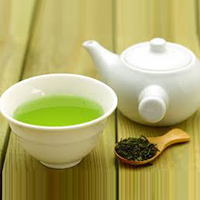
Green tea : The cheerful way to good health

Tea is an infusion of flavorful leaves that has been consumed for centuries as a beverage and is valued for its medicinal properties. Today, scientific research has validated its healthful effects and the cup that cheers has gained recognition as the cup that heals.

The tea shrub (genus Camellia, family Theaceae) is a perennial evergreen with its natural habitat in the tropical and sub tropical forests of the world.  Cultivated varieties are grown widely in its home countries of South and South East Asia, as well as in parts of Africa and the Middle East.  The young shoots or flushes are plucked and processed into green (unfermented), black (fermented), oolong (red, partially fermented) or yellow (partially fermented) teas. In fermented teas, the action of leaf oxidizing enzymes, (mainly aka polyphenol oxidase) convert the tannins and catechins in tea leaves into brown/red colored products1.

Green tea (Camellia sinensis) has  been acclaimed for its antioxidant properties, attributed to the presence of catechins such as epigallocatechin gallate (EGCG). These compounds promote health by preventing lipid oxidation and have been proven to possess antibacterial and antiviral action as well as  anticarcinogenic and antimutagenic properties.
 

Chemistry

The catechins in green tea are responsible for its medicinal properties2.

Other biologically active compounds present in green tea include the methylxanthines, theophylline, theobromine and caffeine. Theophylline has been used as a bronchial smooth muscle relaxant in the treatment of asthma and bronchitis.
 

Biological effects of the catechins in green tea

The biological benefits associated with green tea catechins especially epigallocatechin gallate,  are generally attributed to their antioxidant activity2. They are also reported to scavenge free radical oxygen3.  In studies with lard or vegetable oil, the tea catechins were found to reduce the formation of peroxides more effectively than dl-a-tocopherol or BHA. The antioxidative activity increased in the following order: EC<ECG<EGC<EGCG.  In view of these results, the potential use of the catechins in green tea as effective natural antioxidants in foods has also been explored.

 Tea catechins are believed to act as anticancer agents by detoxifying cancer causing substances in vivo 4,5. In vitro studies revealed that catechin gallates selectively inhibit 5 a-reductase. This enzyme is responsible for the conversion of testosterone to 5-a dihydrotestosterone6. 5-a dihydrotestosterone at high levels, has been implicated in the etiology of prostate cancer and male pattern baldness.
 

Anticancer effects:

The anticarcinogenic effect of green tea extract was studied on mouse skin.  Green tea catechins, especially EGCG inhibited each step in the conversion of a cancer to malignancy. Tea was also found to offer protection against chemically induced tumor initiation, promotion and progression to malignancy as well as inhibit skin cancer induced by ultraviolet radiation7.

In mouse models of chemically induced lung and stomach cancer, there was significant inhibition of tumor incidence and proliferation when green tea catechins were orally administered.  Oral administration of  0.012-1.25% of EGCG or green tea extract to mice or rats with chemically induced oesophegal, intestinal, colon, liver and mammary tumors was found to have a potent inhibitory effect on carcinogenesis.

Recent studies showed that tea drinking reduced the risk of oesophageal cancer in Chinese women; oral cancer in northern Italians; gastric cancer in Swedish adolescents; pancreatic cancer in elderly Poles and residents of a retirement community in the U.S.; and colon cancer amongst retired male self-defense officials in Japan. It is now generally accepted that tea drinking has chemopreventive effects8.
 

Antimutagenic effects:

Green tea catechins have been shown to be antimutagenic, lowering the formation of heterocyclic amines9 (formed during the cooking of meat and fish and proven to be mutagenic). They have also been shown to reduce the occurrence of chromosome aberrations during mutagen exposure10.
 

Protection against atherosclerosis and hypertension:

Lipid peroxidation especially the oxidation of LDL (Low Density Lipoprotein) has been implicated in the etiology of atherosclerosis. In vitro studies confirmed the inhibition of lipid peroxidation induced by cupric ions, by green tea catechins. Green tea extract was found to prevent the increase in serum cholesterol in mice fed high fat diets. A recent cross-sectional study also revealed that in people consuming more than ten cups of green tea per day, there was decrease in serum cholesterol levels, decrease in LDL, VLDL (Very Low Density Lipoprotein) and triglycerides, increase in HDL and reduction in atherogenic index.  In the same study, tea consumption was also found to decrease the levels of  serum markers of  liver damage11. In a study on an elderly group in the Netherlands suffering from coronary heart disease, tea  (probably not green tea) was found to reduce the risk of death from this condition12.

The catechins in green tea were also found to inhibit hypertension in mice (resulting from chronic psychosocial causes) through enhanced sedative action of the brain neurotransmitter GABA 13,  gamma amino benzoic acid. Green tea was also found to lower the incidence of stroke in the elderly14.
 

Protection against infectious diseases

The tea catechins (particularly EGCG and EC) were found to have bactericidal properties. They are believed to damage bacterial membranes. Tea has been used in the treatment of diarrheal diseases and infections such as cholera and typhus15.  Green tea is also believed to have protozoacidal and virucidal16 (including  HIV18)  properties.  However, the effectiveness of tea catechins in the treatment of human viral diseases needs to be confirmed. EGCG has also been shown to stimulate the immune response,  in studies on mice. In this case, the galloyl group in EGCG was postulated to stimulate mouse splenic B-cell proliferation19.
 

Protection against tooth deca

Green tea catechins are believed to offer protection against tooth decay by three mechanisms:

  1. a) By killing the causative bacteria, such as Streptococcus mutans17.
  2. b) By inhibiting the collagenase activity of the bacteria resident below the gum line20
  3. c) By increasing the resistance of tooth enamel to acid induced erosion21


Promotion of longevity

Tea drinking may promote longevity, as evidenced by the low mortality rates amongst Japanese females who are traditional practioners of the tea ceremony22.

Green tea by virtue of its scientifically validated healthful effects has potential utility in the management of a variety of disorders. This ancient herb, associated with wakefulness and harmony in Buddhist legend, has now found its rightful place in modern alternative medicine as a versatile healer.


References

  1. Bokuchava, M.A. and Skobeliva, N.I. (1980). The biochemistry and technology of tea manufacture. Crit. Rev. Food Sci. & Nutr. 12 : 303-370.
  2. Prophylactic functions of tea catechins : Information brochure, Food Research Laboratories, Mutsui Norin Co. Ltd., Japan.
  3. Zhao, B. et al. (1989) Scavenging effect of extracts of green tea and natural antioxidants on active oxygen radicals. Cell Biophysics, 14 : 175-185.
  4. Fujiki, M.D. et al. (1992) Anticarcinogenic effects of (-) epigallocatechin gallate. Preventive Medicine, 21, 503-509.
  5. Bu-Abbas et al. (1995) Stimulation of rat hepatic UDP-glucuronosyl transferase activity following treatment with green tea. Food Chem. Toxicol., 33: 27-30
  6. Shatsung, L., Hiipakka, R.A. (1995). Selective inhibition of steroid 5-a-reductase isoenzymes by tea epicatechin 3-gallate and EGC3-gallate. Biochem. Biophys. Res. Commun. 214 : 833-838.
  7. Mukhtar, H. et al. (1992) Anticarcinogenic effects of (-) epigallocatechin gallate. Preventive Medicine, 21, 351-360.
  8. Schwarz, B. et al. (1994) Coffee, tea and lifestyle. Prev. Med. 23 : 377-384.
  9. Weisburger, J.H.et al. (1994). Prevention of heterocyclic amine formation by tea and tea catechins. Cancer Lett. 83 : 143-147.
  10. Sasaki, Y.F. et al. (1993) The aclastogen-suppressing effects of green tea, Po-lei tea and Rooi bos tea in CHO cells and mice. Mutat. Res. 286 : 221-232.
  11. Imai, K. And Nakachi, K. (1995). Cross-sectional study of the effects of drinking green tea on cardiovascular and liver diseases.Brit. Med. J., 310 : 693-696.
  12. Hertog, M.G. et al. (1993) Dietary antioxidant flavonoids and risk of coronary heart disease: The Zutphen elderly study, Lancet, 342: 1007-1011.
  13. Henry, J.P. and Stephens-Larsen, P. (1984) Reduction of chronic psychosocial hypertension in mice by decaffeinated tea. Hypertension, 6:437-444.
  14. Sato, Y. et al. (1989) Possible contribution of green tea drinking tothe prevention of stroke. Tohuku . Exptal Med. 157 : 337-343.
  15. Shetty, M. et al (19940 Antibacterial activity of tea (Camellia sinensis) and coffee (Coffee arabica) with special reference to Salmonella typhimurium. J. Commun. Dis. 26 : 147-150.
  16. Ryu, E. (1982) Prophylactic effect of tea on pathogenic microorganism infection to animals and humans. Int. J. Zoonoses. 9 : 126-131.
  17. Horiba, N. et al. (1991) A pilot study of Japanese green tea as a medicament: Antibacterial and bactericidal effects. J. Endod. 17: 122-124.
  18. Nakane, H. and Ono,K. (1989) Differential inhibition of HIV-reverse transcriptase and various DNA and RNA polymerases by some catechin derivatives. Nucleic Acids Research, Symposium series 21, 115.
  19. Hu, Z. (1992) Mitogenic activity of (-) epigallocatechin gallate on B-cells and investigation of structure-function relationship. Int. J. Immunopharmacol. 14 : 1399-1407.
  20. Makimura,M. et al. (1993) Inhibitory effect of tea catechins on collagenase activity. J. Periodontol. 64: 630-636.
  21. Yu, H. Et al. (1995) Effects of several tea components on acid resistance of human tooth enamel. J. Dent. 23: 101-105.
  22. Sadakata, S. Et al. (1995). Mortality among female practitioners of Chanyou (Japanese tea ceremony). Tohoku J. Exp. Med. 166:475-477.

Gugulipid | Reduce Cholesterol and ‘Burn’ More Body Fat with This Plant Extract

If you are looking to burn more body fat, gugulipids could be the plant extract for you.

Stroke is the third leading cause of death in North America and Europe, just behind coronary heart disease and cancer. When it happens, it can either be a little one (actually a series of undetected petite strokes over a given period of time) or else, more often than not, one of major consequence that can leave in its wake temporary paralysis and partial amnesia, among other things.

A stroke is initiated with the formation of a blood clot. A clot forms when blood platelets become too sticky and start clustering together like a bunch of grapes. When this happens, the flow of blood starts slowing down in a process similar to 5:00 p.m. traffic backup on the freeway systems of many of America’s major cities.

Gugulipid also has demonstrated in more than two dozen clinical trials, its incredible lipid-lowering properties. It not only lowers total serum cholesterol and plasma triglycerides, but also greatly reduces “bad” cholesterol (LDL-cholesterol), while at the same time increasing “good” cholesterol (HDL-cholesterol). This should come as welcomed news to those who worry about their heart arteries or livers becoming clogged up with too many fat deposits.

Another remarkable accomplishment of Gugulipid is its ability to accelerate the chemical “burning” away of stored fat, by simply converting it into more physical energy. This is done by resetting the body’s own “fat thermostat” a few degrees higher (not to be confused with an unhealthy rise in body temperature). All of this action takes place in the thyroid, that butterfly-shaped gland that sits atop the windpipe at the base of the neck. When Indian scientists administered an isolated ketosteroid from gum guggul to animal models, they noticed, according to their report, “a significant increase in all the thyroid functions” of these rodents. And slightly accelerated thyroid activity with Gugulipid means an increase in the body’s overall metabolic rate, or heightened internal combustion of stored fat.

Gugulipid, in combination with several other plant ingredients of Ayurvedic origin, was studied in humans also. The results of the various formula combinations showed positive effects for weight loss. Gugulipid was the main ingredient in all four combinations studied.

Gugulipid is safer than much of the water we drink or air we breathe these days. It is extremely user-friendly to all body systems and will do our internal vital organs much good if taken in moderate amounts (2 capsules or tablets) each day with a meal of some kind.


Ocimum Sanctum (Holy Basil) To Help With Stress & Immunological Function

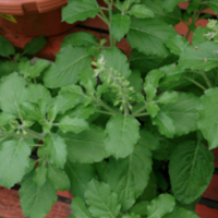
Ocimum sanctum (holy basil), called Tulsi in India, is ubiquitous in Hindu tradition. Perhaps its role as a healing herb was instrumental in its sacred implication. Ayurvedic practice recommends Tulsi in several formulations to enhance immunity and metabolic functions as well as in the management of respiratory problems1.

A variety of biologically active compounds have been isolated from the leaves including ursolic acid, apigenin and  luteolin.  Pharmacological studies have validated the myriad healthful properties of Tulsi. Extracts from the plant have been found to reduce stress, modulate immunological functions and alleviate ulcers in experimental animals2.  Ursolic acid  was found to have anti-allergic properties. When administered to laboratory animals, the compound was found to inhibit  mast cell degranulation and histamine release in  the presence of allergen3.  These studies reveal the potential role of Ocimum sanctum extracts in the management of immunological disorders including allergies and asthma. Recent studies have also revealed the chemopreventive effects of Tulsi extract  in animal models. These effects are mediated through enhanced  carcinogen-metabolizing enzyme activities and raised glutathione levels, facilitated by the active constituents in the extract, such as ursolic acid4.  Researchers have also confirmed the  efficacy of Ocimum sanctum extract in lowering elevated blood sugar levels5.


Phytochemistry

One of the major biologically active compounds in Ocimum sanctum is the triterpene compound ursolic acid.  The structural resemblance of this compound to steroids is believed to be responsible for some of its pharmacological actions3. Ursolic acid is also one of the major constituents of  a well known antioxidant herb, rosemary.

The leaves of Ocimum sanctum yield an essential oil, which contains several biologically active compounds including eugenol, eugenal, carvacrol, mathychavicol, limatrol, and caryophylline. The essential oil has been known to demonstrate antimicrobial activity against Mycobacterium tuberculosis and Staphylococcus aureus in vitro.  Furthermore, a study performed on mice and rats indicated that eugenol and methyleugenol possess adaptogenic (antistress) activity1.

The seeds and mucilage also contain oils with biologically important components.  The seed oil contains fatty acids and sitosterol while the mucilage is made up of xylose and polysaccharides1. The fixed oil of Ocimum sanctum was found to possess anti-inflammatory activity in carrageenan and other mediator-induced paw edema in rats1a.


Clinical Studies

A controlled clinical study validated the role of Ocimum sanctum (Tulsi) as an adjunct in the management of  a  critical metabolic disorder in recent times – diabetes.   A randomized, placebo-controlled  cross-over single blind trial on 40 human volunteers suffering from Type II diabetes was performed.  During the eight week trial, subjects received a daily dose of 2.5 g of Tulsi leaves powder or a placebo for four week periods.  The results of the trial showed a 17.6 % reduction in fasting blood glucose and a 7.3% decline in postprandial blood glucose on treatment with Tulsi as compared to the blood glucose levels during treatment with placebo5

Another study was conducted on rats and mice to observe the anti stress activity of Ocimum sanctum.  To examine this anti stress activity, two groups of 20 mice each were used in a swimming endurance test.  One group was treated with 100 mg/kg of Ocimum sanctum extract while the other group served as the control. The mice were allowed to swim in separate tanks.  The endpoint was noted when each mouse drowned.    The animals treated with Ocimum sanctum showed a significantly  greater physical endurance  than the control group (on an average, the treated animals swam for 1080 minutes as compared to the animals from the control group which swam for 385 minutes).

The effect of Ocimum sanctum extract was explored in another study against ulcers induced by cold stress, restraint ulcers, and aspirin administration in rats.  Cold stress induced ulcers were produced by tying all four of the rat’s limbs and keeping them in a B.O.D. incubator for 2 hours.  Restraint ulcers were created by tying the four legs in a back position.  Aspirin-induced ulcers were produced by the injection of 200mg/(kg of body weight) of aspirin.  In each case, one group served as a control and the other was given Ocimum sanctum extract (100 mg/kg  i.p.).  The stomach of each animal was examined for the presence of ulcers.   After the experiment was complete, it was evident that restraint-stress and chemically induced gastric ulcers were absent in Ocimum sanctum.  The results of these studies indicate that the administration of Ocimum sanctum produces a nonspecific increased resistance against a variety of stress-induced pathological changes in animals2.

The effects of Ocimum sanctum on immunological functions has also been studied. In a study performed on rats immunized by sheep red blood cells (SRBC), it became apparent that pretreatment of Ocimum sanctum enhanced the production of antibodies, including IgE antibody and anti-SRBC.  In addition, the in vitro effect of Ocimum sanctum  on antigen-induced histamine release was studied by sensitizing rats to antigen.  Ocimum sanctum significantly inhibited antigen induced histamine release from mast cells. Based on these results, the authors concluded on the beneficial effects of Ocimum sanctum on  the immune mechanism at various levels, such as on antibody production6.

Experiments also show that Ocimum sanctum possess protective properties against gamma radiation.  The water and aqueous ethanol extracts of Ocimum sanctum were given in single doses and multiple doses before a whole body exposure to gamma radiation in mice.  It was found that the water extract of the plant was more effective and less toxic than the ethanol extract.  Also, an optimum dose of 50 mg/(kg of body weight) of the water extract, at 10 mg/kg of body weight/day for five consecutive days gave the best protection against the effects of radiation7.

In summary, Ocimum sanctum is a safe adjunct to conventional therapy in the management of diabetes and immunological as well as stress-related conditions.  Its record of use in Ayurvedic tradition spans several centuries. Studies performed on animal models to date using levels as high as 100 mg/kg of the extract administered intra-peritoneally, did not reveal untoward side effects2.  Human diabetic subjects receiving 2.5 g of Ocimum sanctum leaves powder  for four weeks also reported  no unfavorable side effects5.

References

  1. Selected Medicinal Plants of India: Chemexcil, 1992, 225-227.
1a. Singh, S. et al. “Evaluation of Anti-inflammatory Potential of the Fixed Oil of Ocimum sanctum and Its Mode of Action”. Journal of Ethnopharmacology, 1996, 54(1), 19-26.
  1. Bhargava, K.P. and Singh, H. “Anti-stress Activity of Ocimum sanctum Linn”.  Indian Journal of Medical Research:  March, 1981, (73) 443-451.
  2. Rajasekaren, M., et. al. “Mast Cell Protective Activity of Ursolic Acid-A Triterpene from the Leaves of Ociumum Sanctum L.  Journal of Drug Development:  1989, 2(3), 179-182.
  3. Banerjee, S., et. al. “Modulatory Influence of Alcoholic Extract of Ocimum Leaves on Carcinogen-Metabolizing Enzyme Activities and Reduced Glutathione Levels in Mouse”.  Nutrition and Cancer:  1996, 25(2), 205-217.
  4. Agrawal, P., et. al. “Randomized Placebo-Controlled, Single Blind Trial of Holy Basil Leaves in Patients with Noninsulin-Dependent Diabetes Mellitus.  International Journal of Clinical Pharmacology & Therapeutics:  Sept. 1996,  34 (9), 406-409.
  5. Bhattacharya, S.K., et. al. “Effect of Ocimum Sanctum Linn. on Humoral Immune Responses”.  Indian Journal of Medical Research:  April, 1988, (87), 384-386.
  6. Devi, P. Uma and Ganasoundari, A.  “Radioprotective Effect of Leaf Extract on Indian Medicinal Plant Ocimum Sanctum”.  Indian Journal of Experimental Biology:  March, 1995, (33) 205-208.

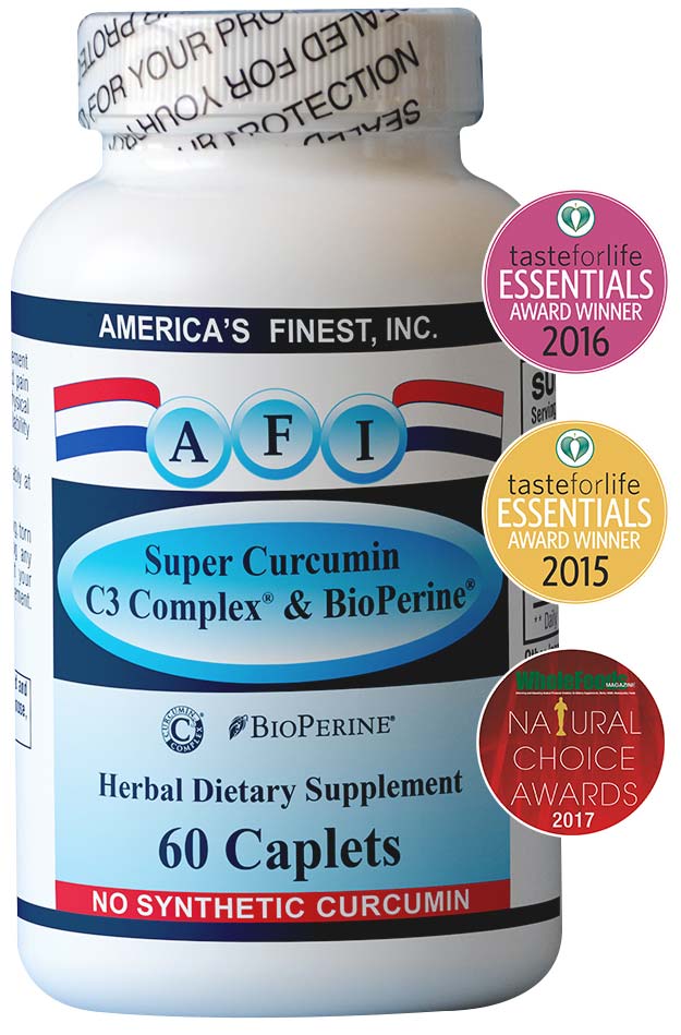
Consumer Alert: Synthetic Curcumin Warning!

Let us face it. Natural product industry is facing its biggest challenge now. On top of the misguided genomic testing, we are now faced with adulteration of natural products especially Curcumin.

As the dietary supplement industry in general and herbal products specifically experience unprecedented levels of negative media coverage and legal action on a regional level, those of us with a long-term commitment to this industry must exhibit leadership. Those of us who have spent our careers making and selling products to improve the health of our fellow human beings know that those writing and saying dietary supplements are either dangerous, unregulated or have no benefit are mistaken, and most of the criticism is unfounded.

While much of the criticism is based upon bad science or lack of knowledge, such as testing extracts with a method no true expert would ever use, those of us immersed in this industry know that there are vulnerable points, particularly in the ingredient supply chain. I believe that Sabinsa is not alone in being dedicated to providing high quality, science-based products to enhance human health and well being, and I call on like-minded companies and the industry’s trade associations to weed out those companies and practices that undermine quality and erode confidence in the entire industry.

We’ve spoken in the past of the importance of respecting and honoring Intellectual Property, which we believe is essential for continued innovation. Recently we have discovered a new threat that must be stamped out to preserve the integrity of the industry and safety of the products we all make.

We have discovered synthetic Curcumin being sold as Turmeric extract with forged Certificates of Analysis. A company selling Curcumin extract in India for export to the US was adulterating their product with 43% synthetic curcumin, but not revealing the synthetic contents. We have taken legal action against them, and a criminal investigation has been opened, but it is important the entire industry be on the lookout for more of this adulteration. I believe it is far more rampant than we thought.

With Curcumin sales in the US alone growing so quickly, it isn’t surprising that there are those looking to sell cheaper, inferior product into the marketplace, but we believe this deceptive practice threatens the future. Our fear is that there are other botanical extracts that are similarly adulterated. We just don’t know, but it’s likely, and the industry needs to find out.

The obvious questions with synthetic herbs are “what was it synthesized from? What chemicals were used, and in what process? How do you know it is safe for consumption by humans?” Synthetically made materials may have distinctively different pharmacological activities compared to natural products. If a company is selling synthetic Curcumin, and not identifying that some or all of it was synthetically derived, that lack of transparency is not only misleading consumers who think they are taking a product derived from Turmeric root, but has the potential to hurt people.

FDA views synthetic versions of natural botanical compounds as different from the botanical itself, thus a supplier of such material would be required to file an NDI notification with FDA, including proof of safety, for the products to be legally sold in the US.

Synthetic copies are, however, difficult to trace in a product using routine analysis. Plant-derived products can be distinguished from synthetic products by their content of natural carbon activity. The use of DNA testing for herbal extracts is debatable, however we know the DNA technique fails when it comes to finding adulteration with synthetic material. Therefore identification and quantification of radiocarbon in these cases provides an accurate way to detect adulteration.

As the industry gets more serious about quality issues, identifying synthetic versions of herbal products becomes crucial. We call on the trade associations and all companies committed to the future of the industry to work together to discover how widespread this deceptive practice is, and to take action to weed it out.

Select the fields to be shown. Others will be hidden. Drag and drop to rearrange the order.
  • Image
  • SKU
  • Rating
  • Price
  • Stock
  • Availability
  • Add to cart
  • Description
  • Content
  • Weight
  • Dimensions
  • Additional information
Click outside to hide the comparison bar
Compare
Shopping cart close